


Photo Source: Nicole Wake
“ Instead of planning surgeries by simply looking at the medical images on a 2D computer screen, surgeons can hold these life-sized physical 3D printed models in their hands and immediately understand the disease.”
From the early anatomical drawings of Leonardo Da Vinci and Vesalius to today, artists,medical students, and professionals have worked to create and perfect accurate representations of the human body. Having access to a precise visualization of anatomical structures is critical to our understanding of body functions, pathologies, and possible cures.

Photo Source: Vesalius Fabrica, Wikimedia Commons
Furthermore, such visualizations are essential maps and tools in surgical planning. The early drawings were created from the dissection of cadavers and this very visual, direct and tactile approach to charting the body’s anatomy is still in use today. However, over the centuries, radiology and more recently digital imaging and 3D scanning have provided non-invasive lenses into the anatomical structure and function. These technologies have also allowed for each individual to obtain maps of his/her body. Today’s #CyantistWeLove, Nicole Wake, a biomedical imaging PhD candidate at the New York University School of Medicine has realized that we can push visualization of anatomical structures one step further and give them life back with 3D Printing. 3D Printed models can provide us with a very tactile and physically meaningful representation of organs of a specific individual, that is much closer to the actual, live organs than 2D and/or virtual imagery. We are excited to feature her and her work at the intersection of medicine, visualization and 3D Printing!
Cyant: You were studying biomedical imaging and had an epiphany: using 3D Printing to convey a more tactile and intuitive feel for organs. What was the spark that gave you the idea to apply your knowledge in medicine and imaging in conjunction with 3D Printing?
Nicole: In my biomedical imaging lab at the New York University School of Medicine, we have a Fortus 360mc printer that was bought to print housing for custom made radiofrequency MRI coils. I was curious to see if we could also use this printer to print anatomically accurate medical models and successfully printed aorta and pelvis models. One day, while carrying one of my aorta models around the hospital, I ran into one of our urologists. He asked me if that was an aorta I was carrying, I said yes, and then he
asked if I could also make kidney tumor models. This led me to test various printing technologies, and I was able to develop a method to create multi-color kidney tumor models.
Cyant: How were you able to create 3D models of organs that can be 3D Printed?
Nicole: In order to create 3D printed medical models from medical imaging data, the 3D medical images have to be segmented, that is, the contours of organs need to be traced very precisely. Then they need to be converted to a file format that the 3D printer understands. Various FDA approved software platforms are available for this and I have become proficient in using several of them.
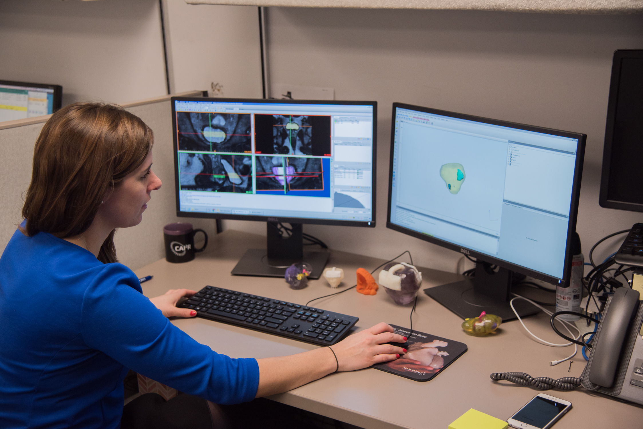
Photo Source: Nicole Wake
Cyant: Once the models are created, which technologies did/do you use, and did you need to create anything new for your purpose?
Nicole: To print multi-color, multi-material 3D printed models, I initially used the Connex500 (Stratasys, Eden Prairie, MN) and most recently have been using the J750 (Stratasys, Eden Prairie, MN). This polyjet technology allows me to print a translucent kidney, along with the other key anatomical structures printed in various colors. The translucency of the 3D-printed models allows easy visualization of the location and size of the tumor as well as the relationship of the cancerous tumor to key anatomical structures such as the renal artery, renal vein, and renal collecting system.
Cyant: So this is opening some really interesting possibilities for medical applications! How is your idea helping physicians plan surgery and obtain new modes of visualization?
Nicole: 3D printed anatomical models help the surgeons to get a whole new perspective of the anatomy of interest. Instead of planning surgeries by simply looking at the medical images on a 2D computer screen, surgeons can hold these life-sized physical 3D printed models in their hands and immediately understand the disease. Patients, medical and/or biology (even high school!) students and professionals alike can better gauge actual dimensions (“My kidney is that big?”) and learn very intuitively from the 3D prints.
Cyant: How are you working with people with diverse skill sets (doctors, technicians) to put your idea to fruition? How receptive were they to your ideas? What have you learned from them in the process?
Nicole: I work closely with our urological surgeons to use 3D printed kidney and prostate tumor models for pre-surgical planning and intra-operative guidance. The urological surgeons believe that these models help them to understand the anatomy and plan procedures. Recently, we performed a retrospective study to determine whether patient-specific 3D printed renal tumor models change pre-operative planning decisions made by urological
surgeons in preparation for complex renal mass surgical procedures. Three
experienced urological surgeons reviewed each renal mass case individually and in a random order to plan an intervention first based on images alone, and again based on images and the 3D printed models. The urological surgeons completed questionnaires about their surgical approach and planning, comparing presumed preoperative approaches with and without the model. In addition, they recorded any differences between the plan and the actual intervention. The results revealed a change in the planned approach in all ten models!

Photo Source: Nicole Wake
Cyant: Given this initial success, what are your next steps and what are you hoping to achieve from here?
Nicole: I am continuing to study the use of 3D printed kidney and prostate tumors in pre-surgical planning. With a fascination for the interface between technology and healthcare, I hope to develop quantitative metrics of how the 3D printed models can make an impact in patient care. We really want to show how these models can be helpful for patients and help them understand and manage their disease.
Cyant: Finally, what words of advice do you have for parents, and young Cyantists, who are inspired by your work and might one day want to work at the intersection of 3D printing and medicine? Are there skills they need to acquire? How can they cultivate their creativity?
Nicole: 3D printing is an enabling technology in medicine and many other fields. Exposing young Cyantists to stories that demonstrate how 3D printing is being used in medicine could help them to develop an interest in the field. In addition, young Cyantists could cultivate their creativity by learning how to use 3D modeling software and the steps required for 3D printing.
Thanks to Nicole for describing her forward thinking work and providing photos illustrating it!